5 Neurology Pearls for the IM, Family Medicine, and PANCE Board Exams
Neurology is a very important part of the Internal Medicine, Family Medicine, and Physician Assistant Board exams and can be challenging at first for many students. In this post, we will review a few key nuggets that will help you analyze many common patient scenarios.
1. In Neurology questions on the medical school clerkship and board exams, nothing helps narrow the diagnosis more than paying attention to the reflexes mentioned in the vignette
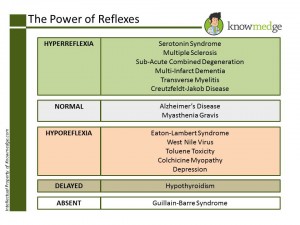
Review the slide below (click to enlarge) for the most high-yield conditions associated with hyperreflexia, normal reflexes, hyporeflexia, and delayed reflexes and absent reflexes.
ABIM Exam Pearls: Reflexes

2. We’re not trained ophthalmologists but remembering these eye conditions can add points to your ABIM score
- Optic nerve lesion → can lead to complete blindness in the ipsilateral eye (monocular blindness of the ipsilateral eye)
- Optic chiasm lesion → Bitemporal hemianopiacommon in pituitary tumors that compress the optic chiasm
- Optic tract lesion → contralateral homonymous hemianopia
- Optic radiation lesion → contralateral homonymous quadrantanopia
In normal individuals, if asked to look the right, the right eye should abduct and the left eye should adduct. If a patient with MS is asked to look to the right (for example), he/she will be able to abduct the right eye but fails to adduct the left eye → Lesion is Left MLF.
Same concept applies when asked to look to the left. Normally, the left eye will abduct and the right eye should adduct. In patients with MS, patients lose the ability to adduct the right eye → Lesion is Right MLF.
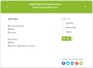
Argyll Robertson Pupil → eyes will be able to constrict when the patient focuses on a near object (eg. bringing fingers to the nose). This is known as accommodation. However, patients with an Argyll Robertson pupil lose the ability to constrict the eyes when bright light is shined into their eyes. In a nutshell, the eyes can’t react to light but can accommodate. This condition is often seen in patients with syphilis.
Here’s a handy mnemonic to remember this feature of Argyll Robertson Pupil:

Marcus Gunn Pupil → This condition is also known as Relative Afferent Pupillary Defect (RAPD). In normal individuals, when a swinging flashlight test is performed, both the direct and consensual eye should constrict to light.
With Marcus Gunn pupil, let’s suppose the left eye is affected. If light is shined into the right eye, both the direct and consensual will constrict. When light is shined into the left eye, both the direct and consensual eye will seem dilated (lack of constriction) → Shows damage to the ipsilateral optic nerve.
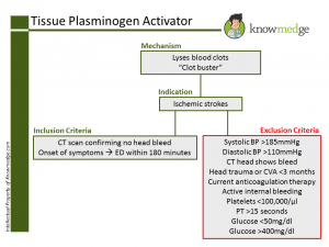
3. Know the indications and contraindications of use of t-PA.
Indications:
- Ischemic stroke as seen on CT head with CLEARLY defined onset of symptoms
- Time of onset of symptoms to administration of t-PA should be no later than 3 hours (180 minutes)
- Blood pressure greater than or equal to 185/110mmHg
- CT head indicates a hemorrhagic stroke rather than an ischemic stroke
- Major trauma to the head within the past 3 months
- Major surgery within the past 14 days
- Current use of anticoagulants as administering t-PA with anticoagulants increases risk of major bleeds
- Platelet count of less than 100,000/uL
- PT>15 seconds
- Glucose 400 mg/dl
Or in slide form…
ABIM Exam Pearls: Tissue Plasminogen Activator (t-PA)

4. Identifying buzzwords is key for selecting the correct neurological diagnosis when CT/MRI findings are included in the vignette.
- Multiple Sclerosis → increased T2 signal and decreased T1 signal. There will be increased enhancement of active lesions with gadolinium.
- Multi-infarct dementia → multiple hypo-dense areas without enhancement.
- Toxoplasmosis, brain abscess, and lymphoma → Ring enhancing lesions seen on CT scan
- Cerebral atrophy → dilated ventricles with dilated sulci
- Normal pressure hydrocephalus → dilated ventricles without dilated sulci. Patient is “wet, wobbly, and weird.” (urinary incontinence, ataxia, and dementia triad is often seen in these patients)
- Alzheimer’s Disease → Brain atrophy with or without periventricular white matter lesions
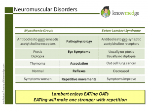
5. Differentiating Myasthenia Gravis and Eaton-Lambert Syndrome can seem challenging at first. That’s why they’re on the ABIM. Ever find yourself second-guessing whether it’s Eaton-Lambert or Myasthenia Gravis that improves with repetitive movements? And, which one is associated with thymoma? Before letting your head spin or do cartwheels, take a few minutes to learn the difference between these two neuromuscular disorders. The concise yet useful categorization will make it difficult to get the two mixed up.
Myasthenia gravis
- Antibodies to post-synaptic acetylcholine receptors
- Ptosis and diplopia can be presenting symptoms
- Can be associated with thymoma
- Reflexes are normal
- Power decreases with repetition
- Antibodies to pre-synaptic acetylcholine receptors
- Ptosis and diplopia are usually absent
- Usually associated with oat cell carcinoma (small cell carcinoma) of the lung
- Reflexes are decreased (hyporeflexia)
- Power improves with repetition (“As you EAT, you get stronger”)
Let’s review it in Knowmedge slide form:
ABIM Exam Pearls: Neuromuscular Disorders

Once again, the folks who write the Internal Medicine med school clerkship shelf and ABIM board exams don’t expect you to have the depth of knowledge regarding neurological conditions that a neurologist possesses. However, topics such as the ones mentioned in the slides and pearls above should assist you with the neurology section of these exams.


[…] […]